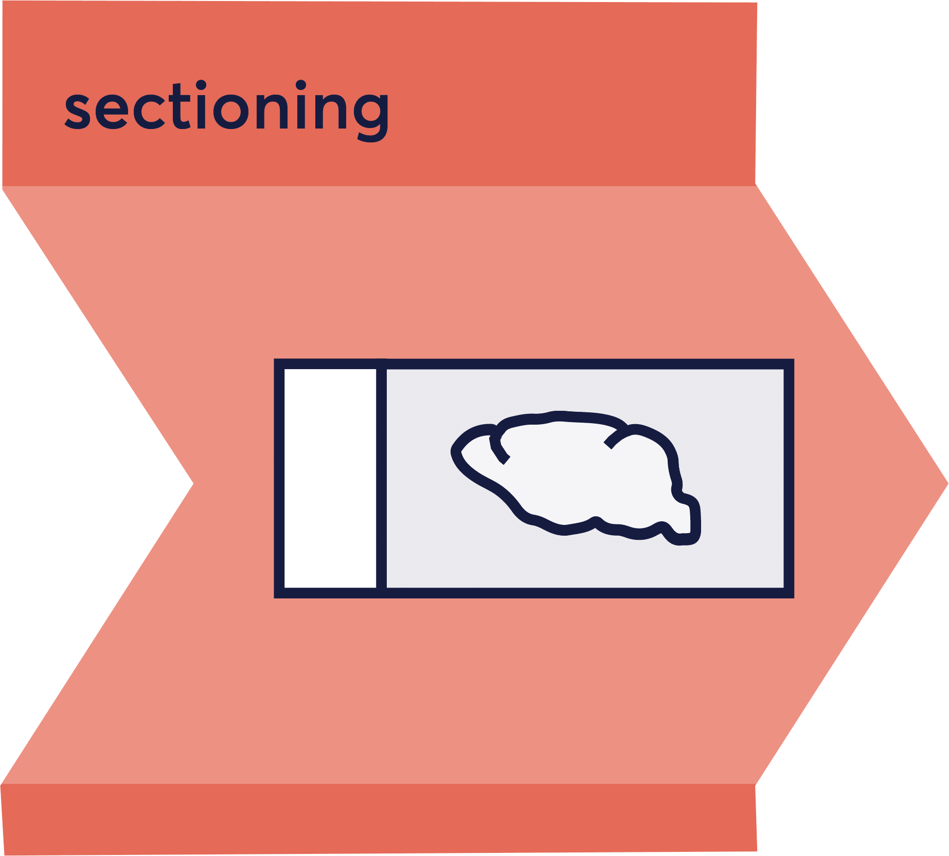
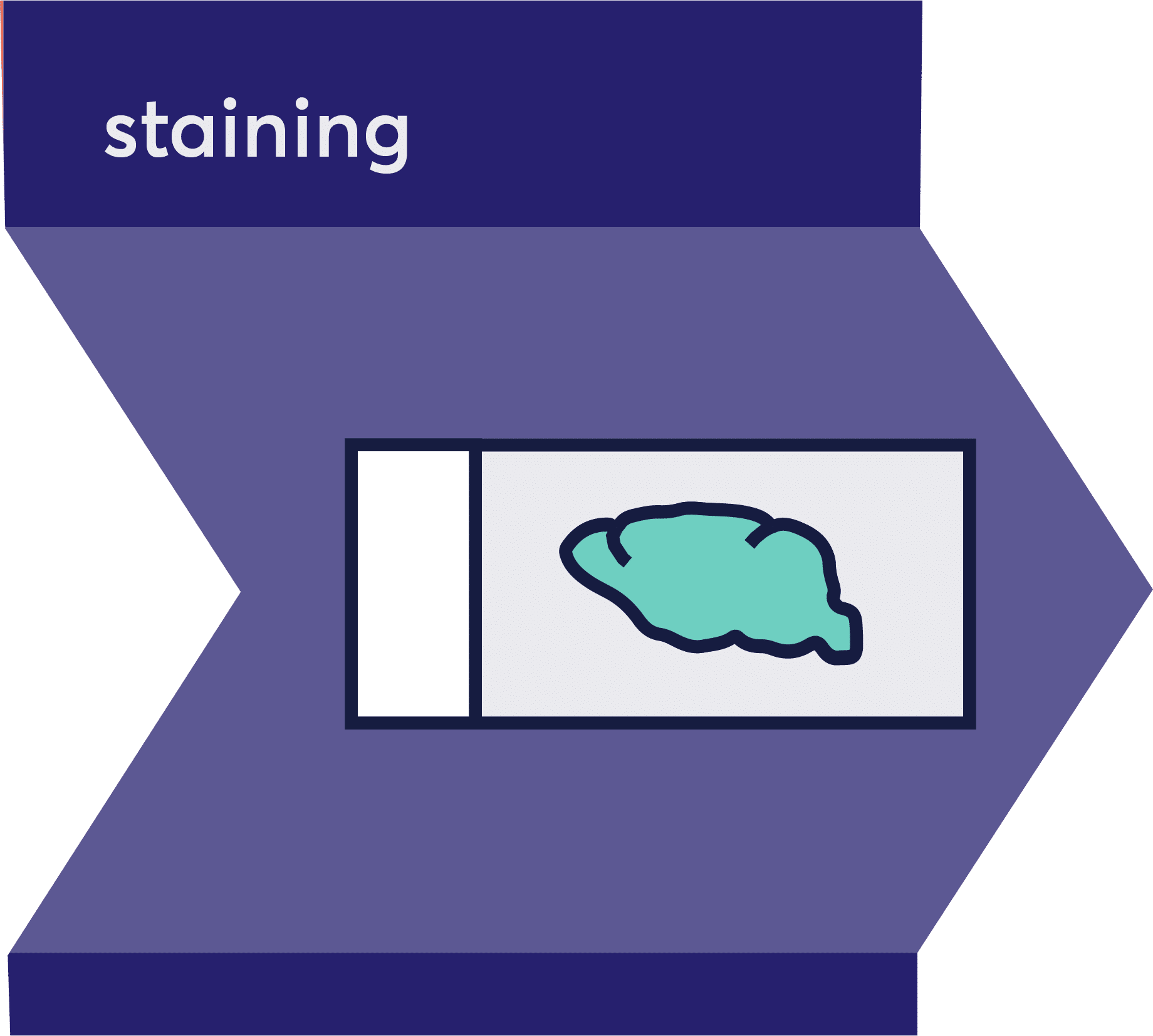
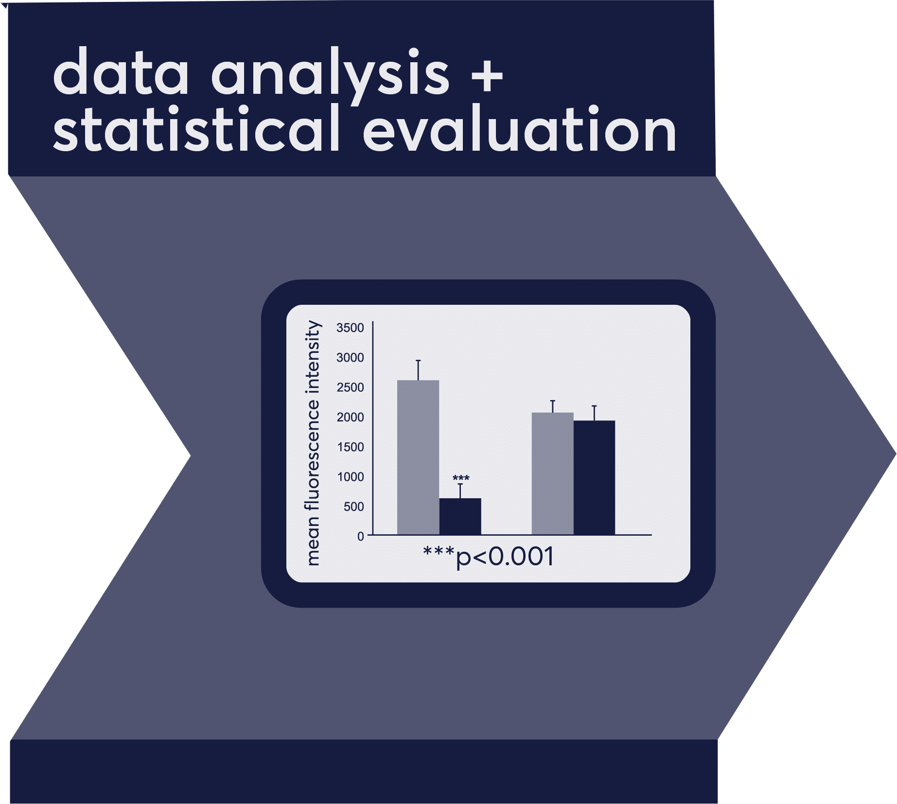
Scantox offers a full set of histology services ranging from collection of tissue samples to delivering study reports that contain all experimental procedures and their outcome. Our approach is based on a sequence of procedural building blocks that allow customizing any service request according to your specific needs. You have the option to select any entry point and exit point in our workflow.
Histology Workflow
Custom-tailored research services for your specific needs

Provide your own animals, take advantage of the large assortment of rodent strains available at Scantox, or ask us to order mice from external sources. Beside rodent samples, we also process human and primate tissue on a standard basis, as well as samples of other species. We are specialized in brain and spinal cord tissue collection but are equally capable of dissecting and analyzing any other tissue of interest, including liver, kidney, spleen lung, muscle, intestines, skin, and other. To maximize the information obtained per tissue sample, Scantox also offers biochemical analyses of parts of the same animal. Sectioning can be fully customized depending on your needs, including parameters such as sectioning plane, section thickness, collection scheme, number and orientation of sections on slides.
Samples are processed using state of the art equipment and well-established protocols. In addition, we constantly establish new markers and reagents for our customers, and our services include literature research, identification and selection of promising markers, comprehensive tests, and validation of the labeling. We have hundreds of established antibodies in stock at any given time, allowing for an almost unlimited number of possible target combinations. We offer 5-channel fluorescence imaging, so tissues can be labeled with up to four different antibodies plus DAPI counterstain of cell nuclei. Automated image acquisition is fast and cost efficient, resulting in high resolution whole slide images. Image analysis is macro-based and runs automated, resulting in fast, unbiased, rater-independent delivery of quantitative data. However, image analysis can be also customized, focusing exactly on the labeling detail you are interested in.
To summarize, we provide fast in-house processing of your valuable samples by experienced and well-trained staff, and we work with reliable shipping companies, to guarantee safe processing and transfer of your biological samples.
Highly standardized default procedures are established for dissecting the target tissue, accurately trim the tissue block, fixation and embedding of your tissue samples. However, customized protocols can be used or established upon request. Just discuss your needs with us, and we take care of it. We are experienced working RNase-free.
Fixation
- Standard: 4% paraformaldehyde
- Alternatives: Glutaraldehyde, zinc, picric acid
Embedding
- Standard: OCT
- Paraffin embedding is done in cooperation with a local external partner
- Choose your preferred orientation (coronal, sagittal, horizontal, longitudinal, transversal)
- Preparation of tissue arrays upon request

Our expertise covers a wide range of sectioning techniques and tissue collection strategies. We process your samples according to the specific requirements of your project.
Sectioning
- Cryotome (Leica, CM3050S and Thermo, NX-70)
- Vibrating blade microtome (Leica, VT1000S)
- Paraffin sectioning is done in cooperation with a local external partner
- Choose any orientation and specify the section thickness
- Uniform systematic random sampling available
(for explanation see: http://www.stereology.info/sampling/)
Collecting
- Specify your collection scheme
- Systematic uniform random sampling available
(For explanation see: http://www.stereology.info/sampling/) - Free floating sections
- Individual or multiple sections mounted on slides
- Tissue arrays
- Choose slides according to your needs

Staining Techniques and Applications
We routinely perform multichannel immunofluorescence (up to 4 antibodies plus DAPI staining in one experiment), using our stock of hundreds of established antibodies. We also constantly establish new markers. Upon request we perform chromogenic immunochemical staining. We are happy to establish and optimize customized staining protocols for your specific needs. While we do not perform in-house pathology services, we cooperate with external partners to offer such analyses in a one-stop shop together with our in-house services.
Our Expertise Includes:
- Hundreds of established antibodies and protocols
- Expertise in processing and analyzing many tissue types of various species
- Antibody testing and optimization
- Target validation of viral infections
- Target validation in transgenic models
- Compound detection
General Stainings
- Hematoxilin-Eosin
- Cresyl violet (Nissl)
- Luxol fast blue
- … and many more
Special Stainings
- Thioflavin S or Congo Red (amyloid plaques, tau tangles)
- Golgi-Cox silver impregnation
- Prussian Blue (microbleedings)
- … other stainings upon request

Multidimensional Image Acquisition
What we offer:
- Automated and comprehensive multidimensional image acquisition
- Mosaic (tiled) high-resolution whole-slide scans for systematic quantifications
- Up to 5-channel epifluorescence imaging (green, red, infrared, far red, and nuclear dye DAPI)

Appropriate Imaging Controls
Autofluorescence is often a neglected, yet serious problem. Most tissues exhibit a certain level of autofluorescence that might be misinterpreted as specific staining. The unspecific signal usually increases with age and with the size of neuronal cells; this is therefore a common nuisance in human brain samples.
To control for this phenomenon, we often devote one of the five available fluorescent channels to image the „intrinsic“ fluorescence. Subtraction of this channel from the „labeled“ channels allows for determination of the specific signal deriving from targeted structures.
In the example, microglia (green arrows) are specifically labeled by Iba1 antibody in the 550 nm channel. Strong signal deriving from either erythrocytes (white arrowheads) or intracellular lipofuscin grains (white arrows) is identified as unspecific autofluorescence because it appears also in the unlabeled channel at 488 nm. Neuronal somata are labeled with NeuN at 650 nm.


Example of image correction using the autofluorescence channel: (A) the original Iba1 image. (B) The autofluorescence channel. (C) The result when B is subtracted from A to remove non-Iba1 background signal. The corrected image can now be used to quantify specific Iba1 signal.
We use sophisticated macro-based software solutions to automatically analyze different features of brain morphology and pathology, e.g., numerical densities of cell somata or axonal fibers, synaptic density, dendritic arborization, area and volume of brain areas or plaques. We are pleased to develop new quantification methods, as well as to assess used (feasibility analysis) or ameliorate existing methods (proof of measurement).

Identification and Detection of Labeled Structures
- Accuracy of detected labeled structures through:
- Validation of antibodies on knockout tissue whenever possible
- Appropriate imaging controls
- Determination and consistent usage of thresholding and filtering parameters throughout the whole study
Analysis of Region and Object Features
We routinely offer a wide variety of automatically generated readouts:
- Total immuno-reactive area
- Sum or mean fluorescence intensity of immuno-reactive particles
- Number, size, density, and shape of labeled particles
- Size exclusions and binning of objects
- Exact morphometric delineation of regions of interest for volume estimation
- And many more
Statistical Data Analysis
- Professional statistical data analysis (unbiased and representative) of behavioral tests and body measurements performed by our team of scientists and biostatisticians
- Conscientious quality checks of measurements and results
Reporting of Experiments and Results
- At the end of each study a full study report including all experimental procedures, qualitative and quantitative assessment of the results, as well a comprehensive statistical analysis will be provided
- Constant updates throughout the study are provided if desired

I would like to express my sincere thanks to all of you for your great work. The professionalism and quality of the planning, implementation and evaluation of the work really impressed me/us! MANY THANKS for your commitment and your really good work.
Thank you very much – I hope that further projects will follow. In any case, we were very impressed by your professionalism and the quality of the data!


