Tau proteins belong to the family of microtubule-associated proteins and play an important role in stabilizing the neuronal microtubules network. They are the major constituents of intraneuronal and glial fibrillar lesions described in Alzheimer’s disease and numerous neurodegenerative disorders referred to as ‘tauopathies’, including progressive supranuclear palsy (PSP), corticobasal degeneration (CBD), Pick’s disease (PiD), argyrophilic grain disease, as well as the inherited frontotemporal dementia and parkinsonism linked to chromosome 17 (FTDP-17).
Molecular analysis revealed that hyperphosphorylation might be the important event leading to tau aggregation resulting in neurodegeneration and dementia. Development of new compounds capable of preventing tau hyperphosphorylation is an increasingly hot topic.
Thus, reliable models are needed that reflect tau hyperphosphorylation in human diseases. For this purpose, we generated the stably transfected SH-SY5Y cell line over-expressing the longest human Tau441 isoform comprising two disease related mutations (V337M/R406W). The phosphorylation pattern of tau in SH-SY5Y-TMHT441 cells can be reliably modulated by several kinase inhibitors. Effects on tau phosphorylation (e.g. at Thr181, Ser202, Thr231/Ser235, Ser262 and Ser396/Ser4049) can be determined by various methods including immunosorbent assay (MSD), immunoblot analysis or mass spectrometry (Löffler T. et al, J Mol Neurosci 2012 May;47(1):192-203).

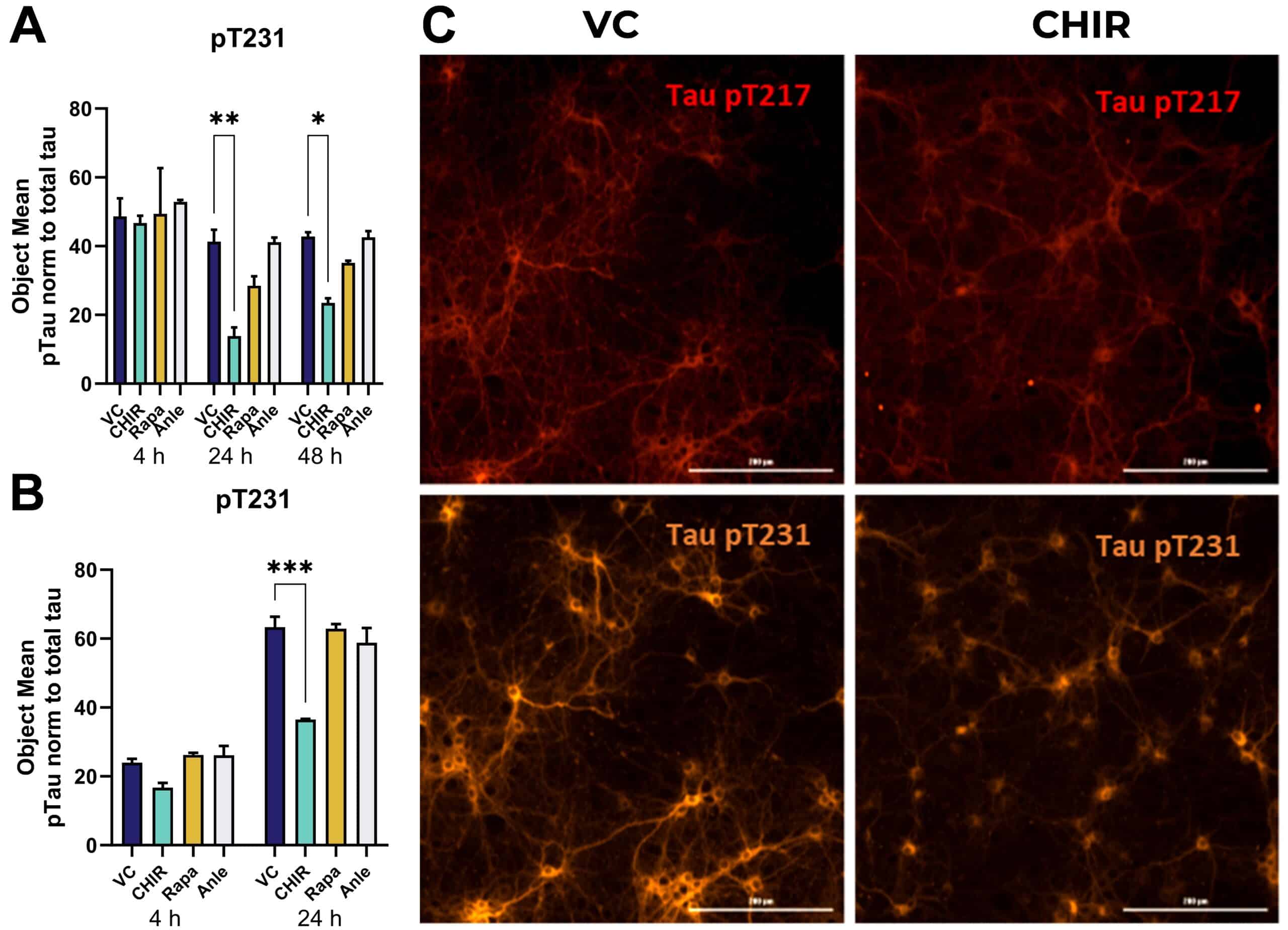
Figure 1: Tau phosphorylation in differentiated stably transfected SH-SY5Y cell line overexpressing 2N4R tau with V337M/R406W mutations after 8 and 24 h of incubation with compounds. Tau phosphorylated at Ser202 as well as Ser396 was assessed with MSD immunosorbent assay in RIPA extracts of the cells, harvested 8 h (A, B) or 24 h (C, D) after treatment with 20 mM LiCl, 1 µM CHIR99021 and 1 µM GSK-3β VIII. One-Way ANOVA with Dunnett’s multiple comparisons test vs. vehicle control (VC). Mean ± SEM; n = 6. **p<0.01; ***p<0.001.
The same assay can also be set up in SH-SY5Y cells over-expressing the longest human Tau441 harbouring P301L mutation or even primary neurons from transgenic mice, such as PS19 mice.


