The balance between neurogenesis and cell death plays a critical role, not only in the embryonic brain development but also for maintenance of the adult brain. Alterations in these processes are observed in many neurodegenerative diseases. The hippocampus, a brain area critical for learning and memory, is especially vulnerable to damage at early stages of Alzheimer’s disease (AD). Emerging evidence indicates that compromised neurogenesis in the adult hippocampus represents an early critical event in the course of AD. For this purpose, Scantox provides a cell culture model to assess effects of your developmental compound on neurogenesis of hippocampal neurons.
Neurogenesis Solution
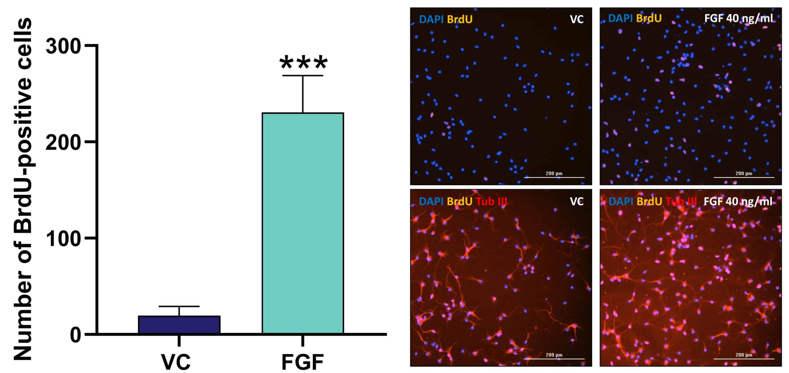
Primary mouse hippocampal neurons are treated with compounds on DIV1 for a total of 48 h. After 24 h, BrdU [10 µM] is added. Compounds are present throughout the assay. On DIV3, after 48 h incubation, cells are subjected to immunofluorescence analysis. Digital images from primary mouse hippocampal neurons are analyzed for the percentage of BrdU-positive cells compared to the total number of neurons (β-tubulin III-positive cells) using Gen05 software (BioTek).

Figure: Effects of fibroblast growth factor (FGF) on neurogenesis of primary mouse hippocampal neurons. Mean + SEM (n=6 / group). Unpaired t-test ***p<0.001. Representative images of DAPI (blue), BrdU (purple) and β-tubulin III (red) positive cells. Scale bar: 200 µM.


