Lipopolysaccharide (LPS) Induced Neuroinflammation
Neuroinflammation is a common feature of several neurodegenerative diseases, such as Alzheimer’s disease, Parkinson’s disease, frontotemporal dementia, amyotrophic lateral sclerosis (ALS), Huntington disease, multiple sclerosis, several lysosomal storage diseases and many more. Recent research suggests that targeting neuroinflammation might present a valid method to treat neurodegenerative disease symptoms. To model neuroinflammation independently from other disease-relevant pathologies, mice can be peripherally injected with the inflammatory stimulus lipopolysaccharide (LPS) to cause an acute neuroinflammatory response. Several different LPS treatment regimens are published and can thus be performed according to your needs.
The most important characteristics of LPS-induced mice are:
- Activated microglia
- Increased inflammation
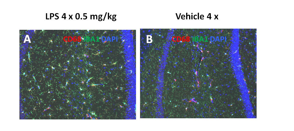
After daily treatment with 0.5 mg/kg LPS for four days, increased microglia activation in the medial hippocampus of LPS-treated mice can be observed (Figure 1).

Figure 1: Immunofluorescent labeling of the medial hippocampus of LPS-treated mice. Animals were intraperitoneally injected with vehicle (A) or 0.5 mg/kg LPS (B) on 4 consecutive days. Tissue was labeled with CD68 antibody (red) for macrophages, IBA1 antibody (green) for microglia and DAPI (blue) for nuclear staining. Samples were collected 24 h after the last injection.
Scantox offers a custom-tailored study design for the induced LPS mouse model, and we are flexible to accommodate to your special interest. We are also happy to advise you and propose study designs. Manifesting microglia activation and an increase in inflammation shortly after treatment grants a remarkably fast processing time for your neuroinflammation study. Furthermore, vehicle-injected mice can serve as control needed for proper study design.
We are happy to evaluate the efficacy of your compound in this induced neuroinflammation animal model! Most important readouts include:
