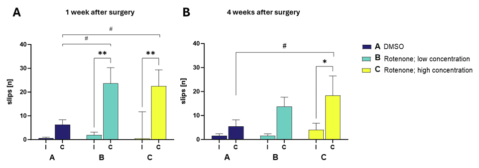
Injection of two different concentrations of rotenone directly into the right striatum of wild-type mice causes a significant increase of contralateral slips in the beam walk test when compared to the ipsilateral side of the same animals as well as the contralateral side of DMSO-injected littermates (Figure 1A). This phenotype is measurable for at least 4 weeks after the lesioning with the high dose of rotenone (Figure 1B).

Figure 1: Number of ipsilateral (I) and contralateral (C) slips in the beam walk test while crossing a 10 mm squared beam 1 (A) and 4 (B) weeks after surgery. Mean + SEM. n=3-6. Two-way ANOVA followed by Bonferroni’s post hoc test; *compared to contralateral control injection; #compared to DMSO injection. **p > 0.01, *p > 0.05.
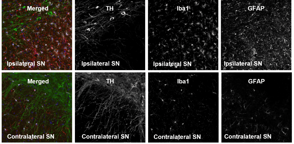
Furthermore, histological evaluation of unilaterally rotenone-injected brains revealed neurodegeneration in the substantia nigra (SN) 4 weeks post administration observed by a decreased tyrosine hydroxylase (TH) signal of dopaminergic neurons (Figure 2). In addition, glial fibrillary acidic protein (GFAP) and ionized calcium-binding adaptor molecule 1 (Iba-1) levels are increased, suggesting astro- as well as microgliosis, respectively (Figure 2).

Figure 2: Representative images of TH, Iba1 and GFAP labeling in the ipsi- and contralateral substantia nigra (SN) 28 days after injection of the high dose of rotenone.
Rotenone is a natural pesticide that inhibits the mitochondrial complex I and has already been shown to induce inflammation and selectively destroy nigrostriatal dopaminergic cells.
The observed motor impairments, the selective loss of dopaminergic neurons, and neuroinflammation in the nigrostriatal pathway observed in this intrastriatal rotenone model closely replicate the human Parkinson’s disease phenotype, making this model a valuable tool to investigate pathological mechanisms of this devastating disease.
Contact us today to get your in vivo study in rotenone-injected mice started.
Check our additional in vitro and in vivo transgenic as well as induced Parkison’s disease models for alternatives.


