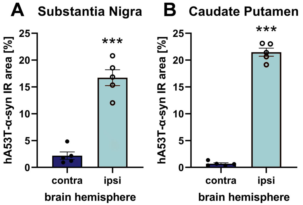
Injecting wild type mice unilaterally with AAV2 hA53T-α-syn virus particles is a fast and at the same time solid method to model Parkinson’s disease brain pathologies. A single unilateral injection of AAV2 hA53T-α-syn into the substantia nigra causes selective expression of hA53T-α-syn in the substantia nigra as well as the caudate putamen of the ipsilateral hemisphere already after 9 weeks (Figure 1).

Figure 1: hA53T-α-syn immunoreactive area (IR) in the substantia nigra (A) and caudate putamen (B) of the contra- and ipsilateral hemisphere after AAV2 hA53T-α-syn injection into the substantia nigra of the ipsilateral hemisphere. Animals were euthanized 9 weeks after injection and brains were evaluated using a human-specific α-syn antibody. n = 5 / group; unpaired t-test; Mean ± SEM. ***p<0.001.
Additionally, activated microglia as labeled by Iba1 as marker of neuroinflammation is increased in the substantia nigra of the AAV2 hA53T-α-syn injected hemisphere. Tyrosine hydroxylase significantly decreased in the AAV2 hA53T-α-syn-injected substantia nigra, indicating a disturbed dopaminergic system in the injected hemisphere (Figure 2).

Figure 2: Iba1 (A) and TH (B) immunoreactive area (IR) in the substantia nigra of the contra- and ipsilateral hemisphere after AAV2 hA53T-α-syn injection into the substantia nigra of the ipsilateral hemisphere. Animals were euthanized 9 weeks after injection. n = 5 / group; unpaired t-test; Mean ± SEM. *p<0.05; **p<0.01. C: Representative images of hA53T-α-syn, tyrosine hydroxylase (TH), Iba1 and DAPI labeling in the contra- and ipsilateral hemisphere of the substantia nigra after AAV2 hA53T-α-syn injection into the substantia nigra of the ipsilateral hemisphere.
Due to the fast onset of disease pathology, this new AAV2 hA53T-α-syn allows a swift processing time of your Parkinson’s disease in vivo study.
Contact us today to get your study in the AAV2 hA53T-α-syn mouse model started!


