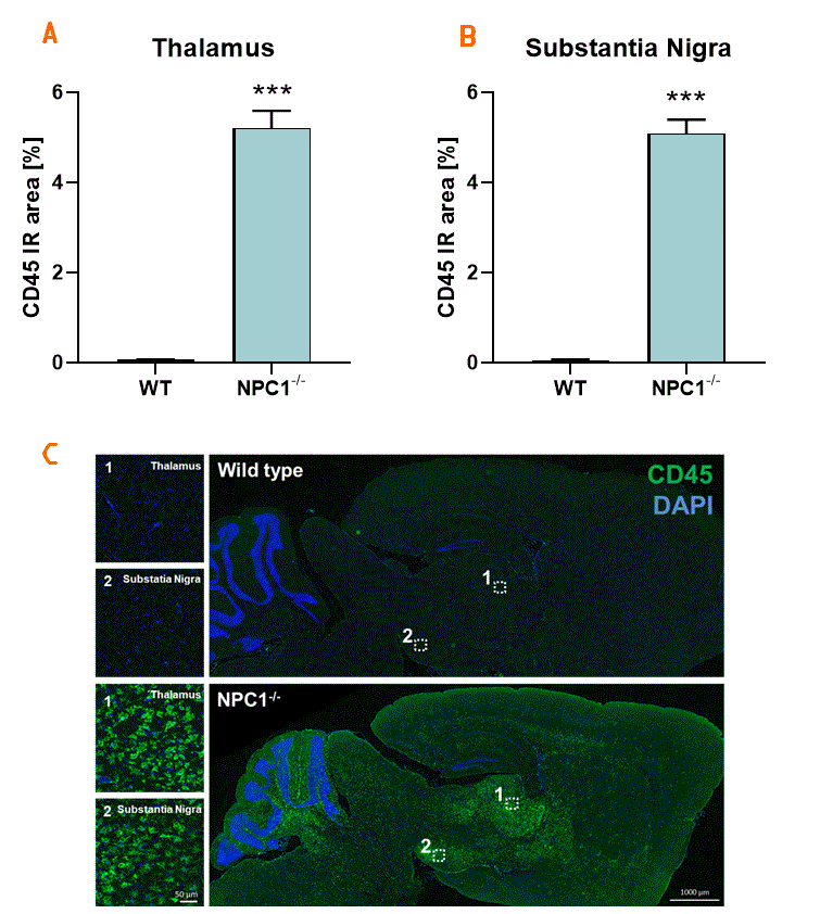
Neuroinflammation is a growing field of research in the fight against neurodegenerative diseases and other disorders affecting the central nervous system. We have previously shown that NPC1-/- mice exhibit strong inflammation (Santiago-Mujica et al., 2019). To further elucidate inflammation in the brain of NPC1-/- mice, we now evaluated CD45 analysis in the thalamus (A) and substantia nigra (B). Our results show that the CD45 immunoreactive area is strongly increased in the thalamus and substantia nigra of 8 week old NPC1-/- mice compared to wild type control animals. Detailed analysis reveals that the density of CD45 labeled cells and the size of these cells is highly increased (data not show).

Figure 1. Quantification of CD45-positive macrophages in the thalamus and substantia nigra of NPC1-/- mice. The thalamus (A) and substantia nigra (B) of 8 week old NPC1-/- mice were investigated for CD45-positive cells of the immune system. Immunoreactive area (IR) in percent compared to control littermates is shown. Unpaired t-test. n = 8 per group. Mean + SEM. ***p<0.001. C: Representative images of CD45 (green) and DAPI (blue) labeling of sagittal brain sections of an 8 week old NPC1-/- and a control mouse.
Contact us today to get your study in the NPC1-/- mouse model started!


