Neurofilament light chain (NF-L) is quantitatively the most common of three different neurofilament chains which constitute the backbone of the neuronal cytoskeleton. When neurodegeneration occurs or axons are damaged, NF-L has been shown to be present in cerebrospinal fluid and even plasma from patients.
The importance of NF-L levels in CSF and plasma as peripheral marker for neurodegeneration is getting more and more attention in humans.
For analysis of plasma and CSF NF-L levels Scantox uses NF-Light® ELISA by UmanDiagnostics, an assay well-described for the analysis of human clinical samples as well as murine samples.
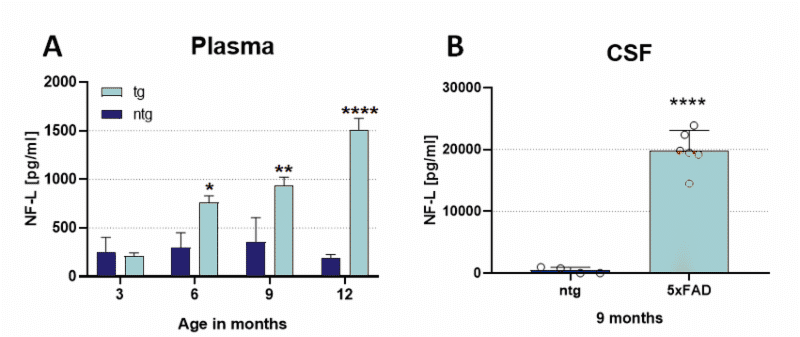
In 5xFAD mice an increase in plasma NF-L levels can be observed already at an age of 6 months, indicating neurodegeneration in this AD mouse model (Fig.1A). Additionally, strongly increased CSF NF-L levels can be observed in 9 months old 5xFAD mice (Fig.1B). Previous analyses using histological quantifications by Oakley et al., 2006 and Jawhar et al., 2012 could validate neurodegeneration in 9 or 12 months old mice, respectively. The analysis of NF-L by NF-Light® thus results in earlier detection of first indications of neurodegeneration and is therefore a well-suited biomarker for early detection of neurodegenerative diseases.

Figure 1: Quantification of neurofilament light chain in plasma and CSF of 5xFAD mice. A: NF-L levels in pg/ml in the plasma of 3, 6, 9 and 12 months old 5xFAD mice compared to non-transgenic littermates. Two-way ANOVA followed by Bonferroni’s post hoc test. B: NF-L levels in pg/ml in the CSF of 9 months old 5xFAD mice compared to non-transgenic littermates. Unpaired t-test. A and B: Mean + SEM. *p<0.05; **p<0.01; ****p<0.0001.
Contact us today to get your your study tissue tested for neurofilament light chain levels!


