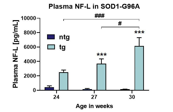
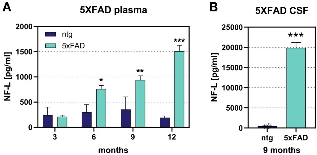
Biomarkers and their use in neurodegenerative disease research delineates a rising area of research and may provide an important link between preclinical disease models and evaluation of disease progression in patients. Neurofilament light chain (NF-L), a neuronal cytoplasmic protein, is increased in a variety of neuronal diseases due to axonal damage. A strong increase of NF-L levels was found in ALS (Amyotrophic Lateral Sclerosis) patients (Gaetani et. al, 2018). Therefore, NF-L levels were investigated in the low copy transgenic mouse model SOD1-G93A. Increased plasma NF-L levels were observed already at an age of 24 weeks, reaching significance with 27 weeks (Fig. 1). In the familial Alzheimer’s disease mouse model 5xFAD an increase in plasma NF-L levels can be observed already at an age of 6 months (Fig. 2A). Additionally, strongly elevated CSF NF-L levels can be detected in 9 month old 5xFAD mice (Fig. 2B).

Figure 1: Quantification of neurofilament light chain in plasma of low copy SOD1-G93A mice. NF-L levels in pg/mL in the plasma of 24, 27 and 30 week old SOD1-G93A mice compared to non-transgenic littermates (ntg). Two-way ANOVA with Tukey’s post hoc test. Mean + SEM. *p<0.05; ***p<0.001. *compared to ntg; # differences between age groups.

Figure 2: Quantification of neurofilament light chain in plasma and CSF of 5xFAD mice. A: NF-L levels in pg/mL in the plasma of 3, 6, 9 and 12 month old 5xFAD mice compared to non-transgenic littermates (ntg). Two-way ANOVA with Bonferroni‘s post hoc test. B: NF-L levels in pg/mL in the CSF of 9 months old 5xFAD mice compared to ntg. Unpaired t-test. A and B: Mean + SEM. *p<0.05; **p<0.01; ***p<0.001.
Contact us today to get your study tissue analyzed for neurofilament light chain levels!


