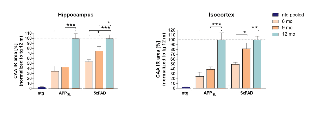
Scantox is your partner for visualization and quantification of Cerebral Amyloid Angiopathy (CAA) in murine tissues. Here we evaluated CAA in APPSL and 5xFAD transgenic animals by measuring the overlap of 6E10 and collagen IV labeling in the isocortex and hippocampus. The strongest progressive increase of CAA signal could be measured in APPSL mice from 6 to 12 months. In 5xFAD mice the total CAA signal was already high at 6 month of age resulting in a weaker signal increase in later age groups. These data suggest, that both mouse models display a progressive and robust vascular CAA pathology, an independent risk factor for cognitive dysfunction of Alzheimer’s disease.
A.

B.

Figure 1: Progression of CAA in different brain areas of APPSL and 5xFAD mice over age. A: Increase of CAA over age in the cerebral cortex and hippocampus measured in overlap with collagen IV labeling expressed as sum immunoreactive (IR) signal normalized to 12 month old transgenic animals. Two-way ANOVA with Bonferroni’s post hoc test, n = 6 per group, females only. *p<0.05; **p<0.01; ***p<0.001. ntg: non-transgenic animal. B: Representative images of amyloid by 6E10 labeling (green) on collagen IV (red) positive vessels (submeningeal arteries) in APPSL mice over age compared to a non-transgenic littermate (ntg). Nuclei are labeled with DAPI (blue).
Contact us today to get your Alzheimer’s disease study tissue analyzed for CAA!


