The importance of microglia for neurodegenerative diseases is well-known and these cells are therefore frequently used as a target for new pharmacological interventions. To study this cell type, isolation of early postnatal microglia from mice is a great tool but does not properly reflect conditions in aged or diseased individuals. Isolation of viable microglia from adult mouse brains of specific disease models via Magnetic Cell Sorting (MACS) thus opens new possibilities to assess the efficacy of microglia-targeting treatments in vitro. Here we investigated the phagocytotic response of isolated adult microglia from 9 months old 5xFAD mice in comparison to age-matched non-transgenic (ntg) microglia. Addition of Aβ1-42 coupled to pH-sensitive pHrodo™ Red label to microglial cells allows to monitor uptake and lysosomal degradation, measurable as increasing red fluorescence in the IncuCyte® Livecell imaging system.
Already 8 h after adding pHrodo™ Red labelled Aβ1-42, 5xFAD microglia show more than doubled fluorescence intensity compared to age-matched ntg microglia, reflecting intense phagocytosis of Aβ in the 5xFAD microglia (Figure 1A). Although also ntg microglia showed phagocytosis of Aβ1-42, this extremely strong phagocytotic response of 5xFAD microglia persisted with time and was at the end confirmed by measuring remaining Aβ1-42 in the supernatant of the cells. 5xFAD microglia took up significantly more Aβ1-42 compared to ntg microglia observable as significantly reduced Aβ1-42 in the supernatant (Figure 1B).
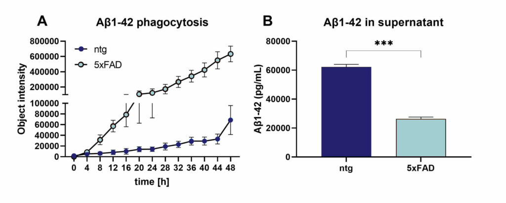
Figure 1: Assessment of Aβ1-42 phagocytosis in isolated adult microglia of 5xFAD and ntg animals. Aβ1-42 phagocytosis was measured for 48 h using pHrodo™ Red labelled Aβ1-42 and IncuCyte® Livecell imaging (A). After 48 h of incubation, the supernatant of the same cells was analyzed for remaining Aβ1-42 (B). n=5 per group. Mean ± SEM. Two-tailed unpaired t-test; ***p<0.001.
Contact us today to get your study in MACS-isolated microglia started!