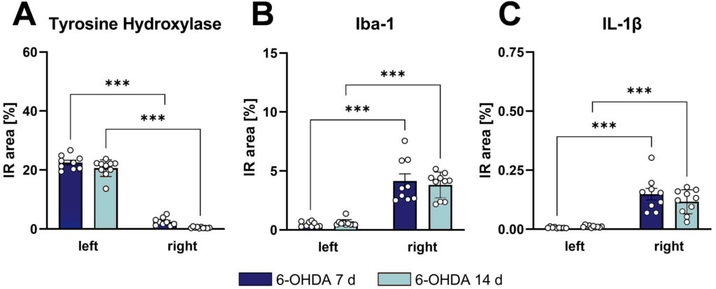
Unilateral 6-hydroxydopamine (6-OHDA) injections in the mouse dorsal striatum (caudate putamen) lead to a strong Parkinson’s disease pathology in the injected brain hemisphere, while the contralateral hemisphere can serve as control. The effect of the 6-OHDA injection can be validated histologically, by measurement of tyrosine hydroxylase (TH) levels in the dorsal striatum that is strongly reduced already 7 and 14 days after 6-OHDA injection (Fig. 1A). In parallel, Iba-1 as marker of activated microglia and IL-1β as marker of the inflammasome are strongly upregulated in the injected brain region already 7 days after injection, indicative for neuroinflammation (Fig. 1B, C).

Figure 1: Effect of a single unilateral injection of 6-OHDA into the murine right dorsal striatum on brain pathology. Wild type mice were injected with 6-OHDA into the right dorsal striatum and the same brain region of both hemispheres analyzed for tyrosine hydroxylase (TH, A), Iba-1 (B), and IL-1β (C) 7 and 14 days after injection. The left dorsal striatum served as control. Two-way ANOVA followed by Bonferroni’s multiple comparison post hoc test; mean + SEM; n = 10 per group; ***p<0.001.
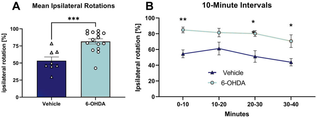
6-OHDA injection into the right dorsal striatum of mice resulted in an increased percentage of ipsilateral rotations compared to vehicle-injected mice in the amphetamine-induced rotation test (Fig. 2). The mean percentage of ipsilateral rotations was highly increased during the whole testing time of 40 minutes (Fig. 2A). Evaluation of rotation behavior in 10-minute intervals showed the strongest effect during the first 10 minutes of testing (Fig. 2B), indicating a highly efficient unilateral lesion of the dopaminergic system in these mice.

Figure 2: Effect of a single unilateral injection of 6-OHDA into the murine right dorsal striatum on lateral behavior in the rotation test. Wild type mice were injected with 6-OHDA or vehicle into the right dorsal striatum and evaluated for unilateral rotations in the amphetamine-induced rotation test 21 days after injection. A: mean ipsilateral rotations in percent relative to total rotations observed during the whole testing time of 40 minutes; B: ipsilateral rotations in percent relative to total rotations observed during 10-minute intervals of the rotation test. Two-way ANOVA followed by Bonferroni’s multiple comparison post hoc test; mean + SEM; vehicle: n = 8; 6-OHDA: n = 16 *p<0.05; **p<0.01; ***p<0.001.
These results show that 6-OHDA injections into the dorsal striatum of mice are a fast method to model Parkinson symptoms that does not depend on genetic mutations compared to transgenic Parkinson’s disease models. Additionally, the 6-OHDA-induced model could easily be combined with a genetic model. The Parkinson’s disease pathology observed in 6-OHDA-injected mice develops within only a few days after injection and can be measured by histological and behavioral analyses.
Contact us today to get your 6-OHDA study started!


