Synapses are crucial contact points between neurons that enable the transmission of information and communication within the nervous system. The structure and function of synapses are generally well understood. But how these synapses form in the first place has long been a mystery. Thanks to a groundbreaking new study recently published in Science, that could be changing. Using CRISPR gene editing technology and human stem cells, the team could observe for the first time the moment that synapses develop in living human cells. What they discovered may change scientists’ understanding of synaptic formation, how proteins reach sites of synaptic formation, and more.
Synaptic Connections Between Neurons
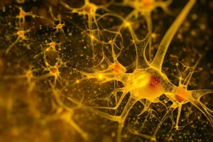
Synapses can be found throughout the entire nervous system, from the brain and spinal cord to muscles and glands. In other words, anywhere that nerve cells are found, synapses are found too. Synapses are essential for the proper functioning of the nervous system and play a critical role in many neurological processes and disorders.
The human brain alone contains over 100 trillion synaptic connections — contact points at which the 100 billion neurons in the brain can connect and communicate. These contact points, or synapses, comprise an axonal terminal, the pre-synapse, and postsynaptic neurons.
How Synapses Form
Detailed in a new study published in Science, researchers from Leibniz-Forschungsinstitut für Molekulare Pharmakologie (FMP) have observed how synapses form in the brain. To accomplish this, the team used CRISPR gene scissors to insert a fluorescent protein into human stem cells and then generated neurons using these modified stem cells. The fluorescent marker allowed the team to observe pre-synapse development as they formed in living nerve cells in real time.
The team’s discoveries are profound and, in some cases, appear to invalidate previous assumptions scientists held about forming pre-synapses.
New Understanding of Pre-Synapses
Pre-synapses comprise three essential elements: scaffolding proteins, calcium channels, and synaptic vesicles. Each element contains its own genes and is made of its own protein molecules. Scientists have long believed that each of the three elements takes its own unique route before joining the other two and forming a functional synapse.
The researchers’ observations tell a different story. As Professor Dr. Volker Haucke, the research group’s leader, explains, “The synaptic vesicle proteins and the proteins of the so-called ‘active zone’ and likely also the adhesion proteins that hold synapses together share the same bus. It was highly controversial. And yet our data in human neurons in culture seem quite clear.”
Furthermore, the team found that axonal transport is mediated by motor proteins known as kinesins. Among these proteins, the primary driver of this process is KIF1A, a motor protein associated with neurological disorders in the peripheral nervous system and the brain. Mutations in KIF1A have been associated with various neurological diseases such as KIF1A-associated neurological disorder (KAND), hereditary spastic paraplegia, and ataxia, suggesting the importance of its role in transporting pre-synaptic proteins.
The researchers also discovered that the axonal transport vesicles responsible for carrying these proteins have a distinct molecular identity. Unlike other vesicles originating from the Golgi apparatus, these transport vesicles share markers with the endolysosomal system. The endolysosomal system is typically involved in degrading defective proteins in non-neuronal cells, so this discovery suggests that neurons have “invented” a unique kind of organelle specifically dedicated to axonal transport.
Looking Forward
A better understanding of the mechanisms involved in axonal transport could have far-reaching clinical applications. Contact points between neurons can be lost or damaged due to aging, injury, or disease. While more research in this area is needed, Dr. Haucke is hopeful.
“Ideally,” he says, “it will be possible to restore or enhance axonal transport to promote neuronal regeneration or counteract aging.”
Scantox is a part of Scantox, a GLP/GCP-compliant contract research organization (CRO) delivering the highest grade of Discovery, Regulatory Toxicology and CMC/Analytical services since 1977. Scantox focuses on preclinical studies related to central nervous system (CNS) diseases, rare diseases, and mental disorders. With highly predictive disease models available on site and unparalleled preclinical experience, Scantox can handle most CNS drug development needs for biopharmaceutical companies of all sizes. For more information about Scantox, visit www.scantox.com.


