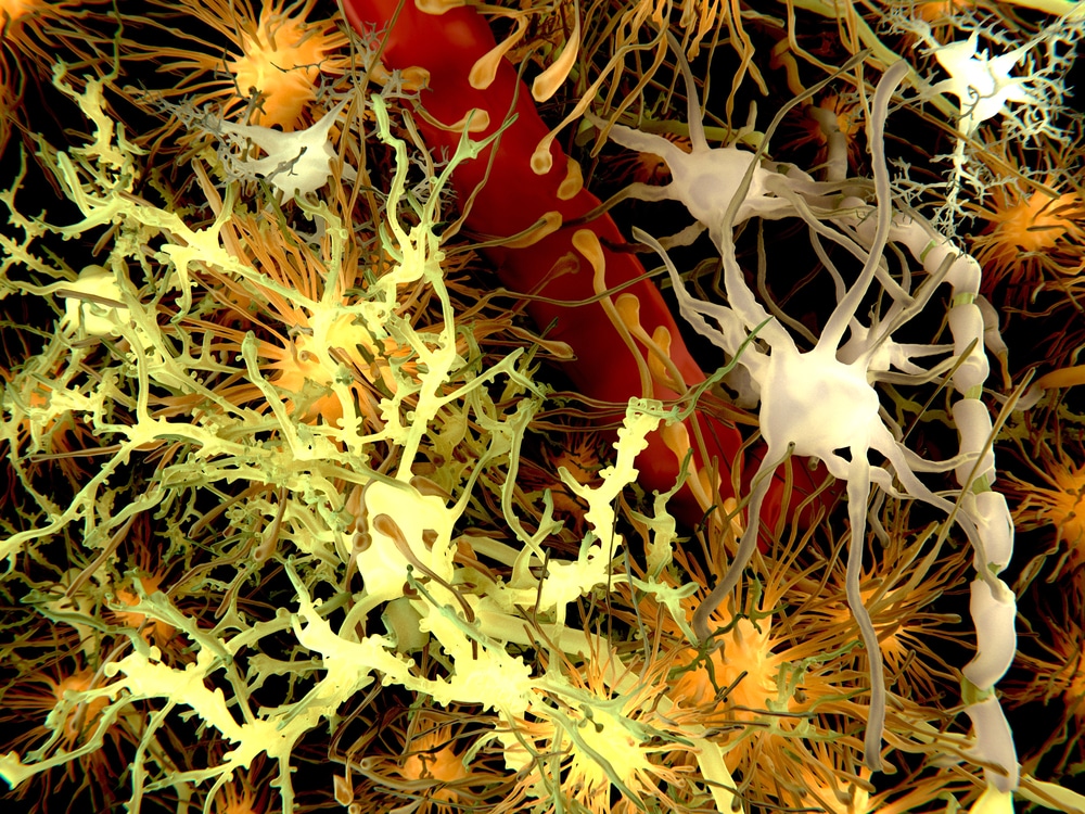
Amyotrophic lateral sclerosis (ALS), also known as motor neuron disease or Lou Gehrig’s Disease, is a degenerative disease that impacts the nervous system. Unfortunately, there are currently no effective treatments, and most ALS patients die within three to five years of diagnosis. As researchers desperately seek treatments for this devastating illness, one type of specialized glial cell comes up again and again: astrocytes. Astrocytes are a key factor in ALS pathology; however, the cells’ exact impact on disease progression has remained a mystery. Now, two new neurological studies conducted by scientists at the Francis Crick Institute may be key to exploring astrocytes’ role in this debilitating disease. Read on to find out more about the techniques used in these studies, including ALS mouse models.

ALS and Astrocytes in Mouse Models and Humans
Biomedical researchers at the Francis Crick Institute published a series of two papers exploring astrocytes’ role in ALS. In the first paper, published in Genome Research, the researchers assessed all existing public datasets of astrocytes in ALS. Those datasets included both human models and ALS mouse models. The researchers found that, in ALS, astrocytes exhibit two key changes. First, they become pro-inflammatory, which is harmful to neighboring motor neurons. Second, they lose important protective functions, including the ability to uptake glutamate, an amino acid that is ordinarily metabolized in the body. Over time, the eventual glutamate build-up damages motor neurons.
What Drives Key Changes in Astrocytes?
After the datasets revealed harmful changes in ALS astrocytes, the researchers needed to know: What, exactly, drives those changes? That question was the crux of the second study, published in Brain. In that study, the researchers found that astrocytes with different ALS-causing genetic mutations also have distinct underlying molecular patterns. In other words, during ALS, astrocytes likely acquire mutation-dependent changes over time. The team continued by examining the impact of different ALS-causing mutations on astrocytes. They found that, even without the impact of neighboring immune cells, these mutations appear to drive harmful changes in the astrocytes. “The nature and diversity of astrocyte transformation between ALS mutations was not well known,” explained study lead Doaa Taha. “The insight we’ve gained into the different ways this change manifests in early disease could be a helpful starting point in efforts to reverse the cell transformation.”
Implications of the ALS Astrocyte Study
“Reversing the cell transformation,” as Taha put it, certainly sounds promising. However, the researchers have more work to do before they can understand the molecular patterns in affected astrocytes. Ben Clarke, co-lead author on both papers, said, “We’re now working to understand the biology of the distinct molecular patterns we’re observing and how they converge. Understanding the early astrocyte changes in ALS could provide us with new therapeutic targets.”
_____
Ultimately, reducing or reversing the changes to astrocytes could prove key in treating ALS. As study lead Oliver Ziff puts it, “These cells are not just innocent bystanders but actively contribute to the progression of the disease.” Though further research is needed, this is one scenario in which assessing both human models and ALS mouse models can prove illuminating.
Scantox is a part of Scantox, a GLP/GCP-compliant contract research organization (CRO) delivering the highest grade of Discovery, Regulatory Toxicology and CMC/Analytical services since 1977. Scantox focuses on preclinical studies related to central nervous system (CNS) diseases, rare diseases, and mental disorders. With highly predictive disease models available on site and unparalleled preclinical experience, Scantox can handle most CNS drug development needs for biopharmaceutical companies of all sizes. For more information about Scantox, visit www.scantox.com.


