Choosing the proper readouts is crucial for every cell culture experiment. Scantox offers a wide range of analyses for different parameters to characterize and quantify effects of test substances.
Our established cell-based assays cover a wide range of parameters including cell viability, proliferation, apoptosis, oxidative stress, mitochondrial activity and cellular signaling pathways, among others. We can also support the process of finding the perfect solution for your screening assay using a customized cellular imaging and analysis assay.
For live cell analysis, we utilize the advanced Sartorius IncuCyte platform. This system enables real-time monitoring and analysis of cellular behavior, allowing for dynamic insights into cell physiology, proliferation, migration, and more. With the IncuCyte, cellular processes in a non-invasive manner can be captured and analyzed, providing valuable information for drug discovery and mechanistic studies. At Scantox, the following assays for neuropharmacological applications are established:
Video: Live monitoring of neurite outgrowth in iPSC-derived neurons with Sartorius IncuCyte platform over 4 days.
Despite cell-based and live-cell assays, samples derived from in vitro treatment studies can also be analyzed for protein or mRNA expression levels to investigate target regulation.
To streamline and enhance the western blotting process, we employ the Simple Western WES platform that allows for high-throughput protein analysis, precise quantification, and reproducible results, saving time and reducing variability in protein analysis workflows.
In addition, we provide analyses using the Mesoscale Discovery (MSD) MESO QuickPlex platform for advanced immunoassays. This highly sensitive and reliable system enables multiplex protein detection and quantification, providing comprehensive insights into protein biomarkers.
Established assays include, but are not limited to:
- MTT
- LDH
- IncuCyte live/dead staining
- ATP assay
- Necrosis (Propidium iodide)
- Apoptosis (YOPRO, Caspase-3 activity, Annexin V)
- TMRM
- Mitotracker
- JC1
- CellROX
- DCFDA
- MitoSOX
- Aβ1-38, Aβ1-40, Aβ1-42, sAPP-α, sAPP-β
- Tau and phosphorylated tau profile
- Signal transduction molecules
- Inflammation markers, cytokine secretion
- Neuronal markers (β-III tubulin, neurofilament, MAP2, tyrosine hydroxylase)
- Glial markers (GFAP, CD11b)
- Disease-related markers (tau, phosphorylated tau, signaling molecules, synuclein)
- Synaptic proteins (synaptophysin)

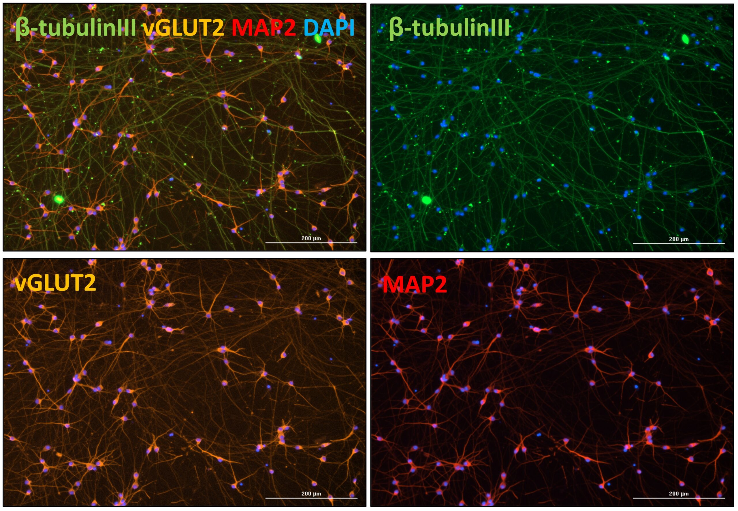
Figure 1: iPSC-derived glutamatergic neurons: Confirmation of maturation to glutamatergic neurons. Cells were fixed on DIV11 and immunocytochemically stained for the neuronal marker β-tubulin III, vGLUT2 and MAP2. Scale bar 200 µM.
- Synaptic proteins (glutamate receptor 1, NMDA receptors, synaptophysin, drebrin) e.g., from matured hippocampal neurons
- Tau and phosphorylated tau isoforms
- Aβ peptide profile, Aβ aggregation assays
- Cell signaling molecules
- TDP43
- Synuclein

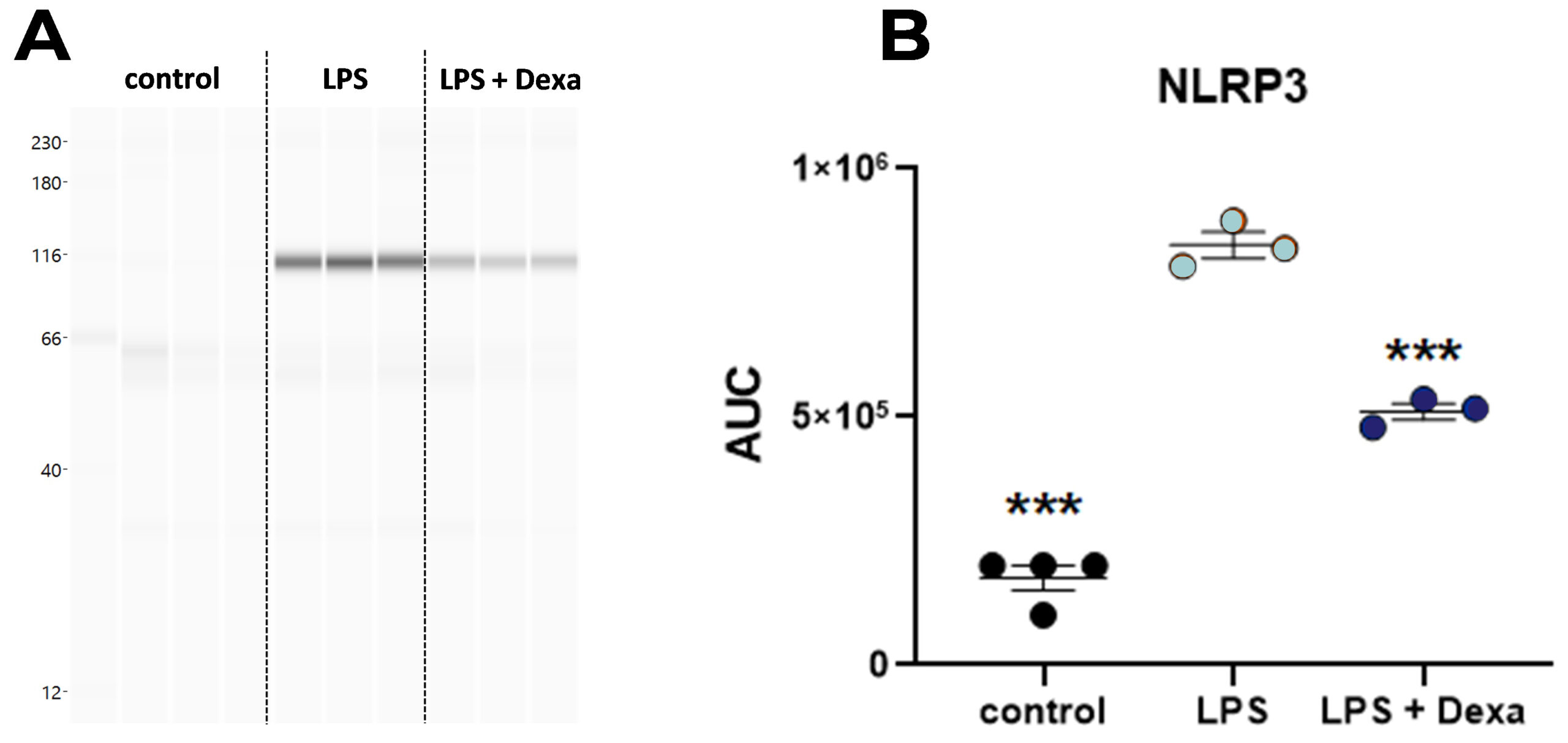
Figure 2: Quantification of NLRP3 in mouse hippocampal slices after LPS stimulation for 24 h. A: NLRP3 quantification of LPS-stimulated organotypic brain slices by an automated Western Blot system (WES). B: NLRP3 area under the curve (AUC) values of LPS-stimulated organotypic brain slices as measured by WES. Aligned dot blot; Mean ± SEM; n = 3 – 4 per group. Dexamethasone (Dexa) served as reference compound. One-way ANOVA followed by Dunnett’s Multiple Comparison post hoc test compared to the LPS-treated group; ***p<0.001.


