Neuroinflammation is a growing field of research in the fight against neurodegenerative diseases and other disorders affecting the central nervous system. We have already shown that 4L/PS-NA mice exhibit strong inflammation in visceral organs and cortical brain regions (Schiffer et al., 2020). Since the cerebellum of 4L/PS-NA mice presents with a highly disturbed morphology (Schiffer et al., 2020), we now evaluated neuroinflammation in this brain region. Our results show that astrocytosis and microgliosis are strongly increased in the cerebellum of 18 week old 4L/PS-NA when labeled with GFAP and IBA1 antibodies, respectively.
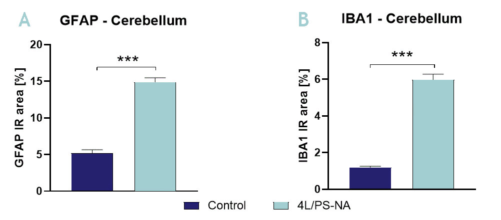
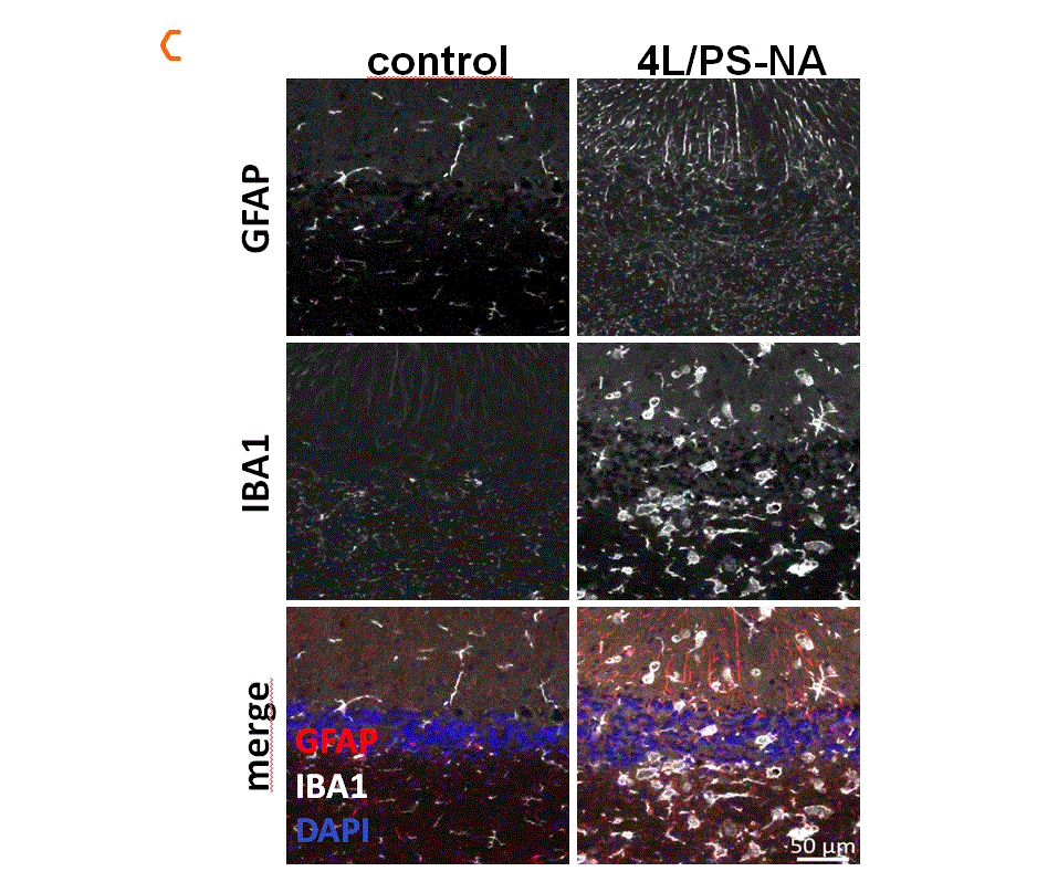
Figure 1. Quantification of astrocytosis and activated microglia in the cerebellum of 4L/PS-NA mice. The cerebellum of 4L/PS-NA mice was analyzed for astrocytosis (GFAP; A) and activated microglia (IBA1; B) immunoreactive area (IR) in percent at the age of 18 weeks compared to control littermates. Unpaired T-test. n = 5 per group. Mean + SEM. ***p<0.001. C: Representative images of GFAP, IBA1 and DAPI labeling of the cerebellum in 18 week old 4L/PS-NA and control mice.
Contact us today to get your study in the 4L/PS-NA mouse model started!


