Besides transgenic rodent models Scantox provides one of the most frequently used animal models for PD utilizing unilateral injection of 6- hydroxydopamine (6-OHDA) into the medial forebrain bundle (MFB) in rats, which results in total denervation of the dopaminergic nigrostriatal pathway.

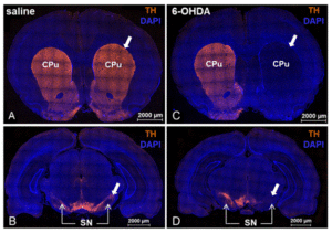
Figure 1: Unilateral injection into the MFB of saline (A, B) or 6-OHDA (C, D) in male Wistar Han rats. Immunofluorescent labeling on coronal brain sections revealed an almost complete loss of tyrosine hydroxylase (TH; orange) in the ipsilateral caudate putamen (CPu; C as well as in the substantia nigra (SN; D) after 6-OHDA injection. Saline injection did not affect TH immunoreactivity (A, B). Cell nuclei were labeled with DAPI (blue). Arrows in A and C indicate CPu, arrows in B and D indicate SN.
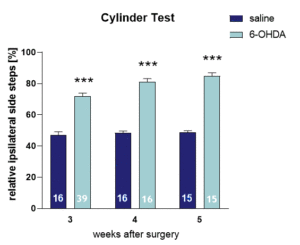
 Figure 2: Cylinder test of 6-OHDA lesioned rats. Ipsilateral fore limb use after unilateral 6-OHDA injection into the MFB. 6-OHDA significantly increased the frequency of ipsilateral fore limb use compared to saline injected control animals 3, 4 and 5 weeks after surgery. Number in columns gives group size (n); mean + SEM; mixed effects analysis with Bonferroni´s post hoc test; ***p<0.001.

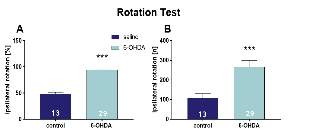
Figure 3: Amphetamine-induced ipsilateral rotation of 6-OHDA-injected rat MFB. Rotational response to D-amphetamine 3 weeks after 6-OHDA injection. 2.5 mg/kg D-amphetamine was injected i.p. on the test day. 30 minutes later, rotations were analyzed in the rotometer bowl. 6-OHDA-injected rats performed almost only ipsilateral rotations while saline-injected rats performed approximately the same number of ipsi- and contralateral rotations (A). The total number of ipsilateral rotations during a 60 min period was significantly increased in 6-OHDA-injected rats compared to saline-injected control rats (B); Number in columns gives group size (n); mean + SEM, Mann Whitney test: ***p<0.001.
Contact us today to get your study in the 6-OHDA-induced rat model started!


