Different preformed recombinant human α-synuclein (α-syn) fibrils were evaluated for their toxicity and seeding properties on primary cortical neurons in vitro.
Our results show that monomeric human α-syn has no impact on cell viability, while preformed wild type and A53T human α-syn fibrils have toxic effects on primary cortical neurons. This toxic effect was exceeded by oligomeric isoforms (Fig.1A). Only A53T α-syn fibrils of the tested isotypes showed seeding properties in cortical neurons (Fig.1B).
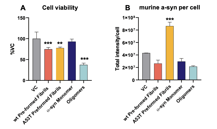
Figure 1. In vitro assessment of α-syn preformed fibrils on mouse primary neurons. Toxicity (A) as well as seeding (B) properties of different preformed recombinant human α-syn species (Stressmarq) on mouse primary cortical neurons. (A) Neurons treated with α-syn species, and assessed for cell viability by MTT assay. (B) Neurons treated with α-syn species and immunocytochemically analyzed for murine α-syn. Mean+SD. One-way ANOVA with Bonferroni post hoc test (vs vehicle control: VC). ** p<0.01, *** p<0.001.
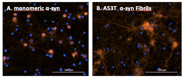
Figure 2. Representative images of endogenous murine α-syn accumulation after seeding with different preformed recombinant human α-syn species (Stressmarq). Neurons were treated with (A) monomeric and (B) A53T preformed fibrils and after incubation immunocytochemically stained for murine α-syn. Nuclear stain DAPI = blue; murine α-syn = red; scale bar 100 µM.
In vivo mouse model expressing α-synuclein with A53T mutation
hA53Ttg mice express A53T mutant human α-synuclein under the control of the murine Thy-1 promoter. This line M53 is bred on a C57BL/6J background (Chandra et al.2005).
Animals show the following phenotype:
- 10 – 20 -fold higher α-synuclein levels compared to wild type mice (Chandra et al.,2005)
- Motor deficits in the RotaRod test at 6 months (Chandra et al., 2005; Rothman et al., 2013)
- Muscle weakness in the Wire Hanging test at ~ 8 weeks (Maki et al., 2019)
- Reduced anxiety in the Open Field and Elevated Plus Maze test starting at 10 and 14 weeks, respectively (Rothman et al., 2013)
- Mean survival of about 43 weeks (Chandra et al., 2005)
The phenotype described above closely reflects PD pathology making the hA53T-Sud mouse a perfect model for your drug testing.
Contact us today to get your study in primary cortical neurons or hA53Ttg mice started!


