Detection of mRNA by fluorescent in situ hybridization (FISH) is a great alternative for proteins that are hard to detect by immunofluorescence. FISH is also the method of choice for the analysis of expression efficacy during the development of new gene therapies.
The Department of Histology of Scantox therefore established the FISH labeling method and is now ready to offer this method to be used for your project. Experiments can be executed on regular cryosections. Due to our vast experience with multichannel immunofluorescence labeling of tissue sections, offering a stock of hundreds of tested antibodies against a large variety of targets, a combination of FISH with immunofluorescent labeling of protein targets can be performed. Up to four different markers plus DAPI staining of cellular nuclei can be used in one experiment.
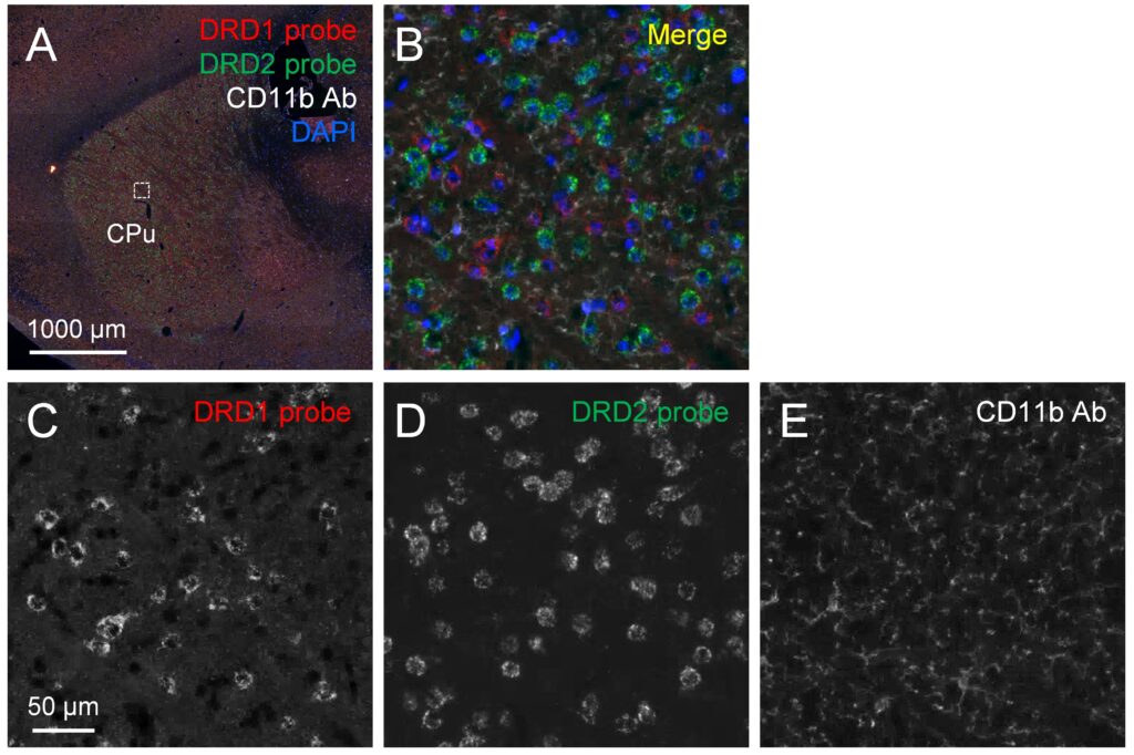
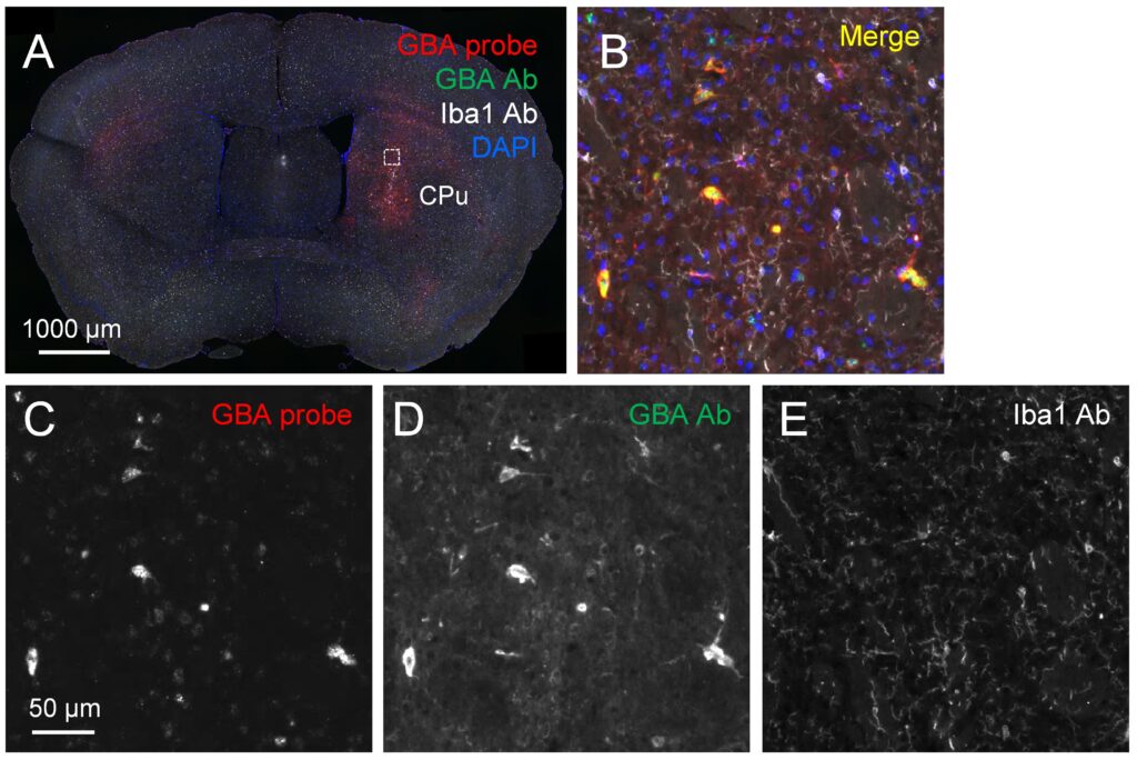
Currently, FISH probes against dopamine receptors D1 (Fig.1C) and D2 (Fig. 1D; D1R and D2R, respectively) as markers of Parkinson’s disease, acid α-glucosidase (GAA) as marker of Pompe disease, glucosyl ceramidase β (GBA, Fig. 2C) as marker of Gaucher disease as well as parvalbumin and green fluorescent protein (GFP) are established.

Figure 1: Dopamine D1 (C) and D2 (D) receptors were detected on different populations of medium spiny neurons in the caudate putamen (CPu) by double-FISH (red and green channels, respectively). FISH was combined with immunofluorescence of microglia using CD11b antibody (E, white channel). The small rectangle in the overview (A) shows where magnified images were taken (B-E).
Our FISH protocol can readily be combined with immunofluorescence as indicated by additional successful antibody labeling for microglia markers CD11b (Fig.1E) and Iba1(Fig. 2E), astrocyte marker GFAP, choline acetyl transferase (ChAT), tyrosine hydroxylase (TH), and GBA1 (Fig.2D).

Figure 2: FISH labeling of GBA in a murine striatal coronal section unilaterally injected with AAV-GBA. GBA probe (C, red channel) is detected in the right striatum around the injection site. Magnification shows FISH-labeling in infected cells, and high protein levels detected by GBA antibody (D, green channel) in the same cells. Microglia are labeled by Iba1 antibody (E, white channel). The small rectangle in the overview shows where magnified images (B-E) were taken.
We welcome your inquiry, and we are happy to discuss collaboration discounts in case control data can be used for marketing purposes by Scantox.


