Parkinson’s disease (PD) is a complex neurodegenerative disorder. Apart from genetic factors, exposure to certain environmental toxins has been associated with an increased risk for developing PD.
By utilizing these neurotoxic agents such as 6-OHDA, rotenone, and MPP+ the cellular damage observed in PD can be closely mimicked. These agents selectively induce oxidative stress and mitochondrial dysfunction, leading to dopaminergic neuron degeneration similar to what is observed in PD patients.
Using these models, the efficacy of potential treatments by examining their effects on neuronal survival and apoptosis can be evaluated.
These assays can be performed on a variety of cell types, including
- SH-SY5Y cells, human neuroblastoma cells often used as a model for dopaminergic neurons
- Primary rodent cortical or hippocampal neurons, offering insights into general neuronal responses (Figure 1 and 2)
- Primary rodent neurons from the ventral mesencephalon, focusing on dopaminergic neurons directly affected by PD (Figure 3)
- iPSC-derived neurons, induced pluripotent stem cell-derived neurons providing a human-specific context and essential for translating findings into clinical applications
When utilizing primary rodent neurons from the ventral mesencephalon, immunocytochemical evaluations are regularly performed to closely investigate and monitor cell health and survival of lesioned cells, focusing on dopaminergic neurons (Figure 3). The following stainings are used as standard readouts:
- MAP2 to assess the overall integrity and structure of neurons
- Tyrosine hydroxylase (TH) to assess dopaminergic neurons critically affected in PD
By employing these lesion-induced models, the pathological environment of PD can be mimicked in specific cell types. This approach allows to identify potential therapeutic agents and accurately monitor the effects of experimental compounds that may protect against or reverse neuronal damage. Furthermore, we are happy to implement new cell culture models, readouts, or customized assays into your next study.
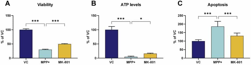
Figure 1: MPP+ lesion on primary cortical mouse neurons. Primary cortical mouse neurons were lesioned with MPP+ and cell viability was assessed by MTT assay (A), level of ATP within the cell by the luminescent ATP assay (B) and apoptosis by YO-PRO-1 assay (C). Data are displayed as percent of vehicle control (VC) and presented as bar graphs with standard error of mean (SEM; n=6-9 per group). MK-801 served as a reference item. For statistical analysis One-way ANOVA followed by Dunnett’s post hoc test versus MPP+ lesion was used. *p<0.05, ***p<0.001.

Figure 2: Rotenone lesion on primary cortical mouse neurons. Primary cortical mouse neurons were lesioned with rotenone and cell viability (A) and cytotoxicity (B) was detected by MTT and LDH assay, respectively. Data are displayed as percent of vehicle control (VC) and presented as bar graphs with standard error of mean (SEM; n=6 per group). MK-801 served as a reference item. For statistical analysis One-way ANOVA followed by Dunnett’s post hoc test versus rotenone lesion was used. ***p<0.001.

Figure 3: MPP+ lesion on primary mouse neurons from the ventral mesencephalon (TH neurons). A: Percent of TH-positive neurons of MAP2-positive neurons. Data are presented as bar graphs with standard error of the mean (SEM; n=6 per group). MK-801 served as reference item. For statistical analysis One-way ANOVA followed by Dunnett’s post hoc test versus MPP+ lesion was used. *p<0.05, **p<0.01. B: Representative images of vehicle-treated cells. Red: MAP2 staining, yellow: TH staining. C: Representative image of vehicle-treated cells. Yellow: TH staining.
We are happy to evaluate the efficacy of your compound in these in vitro PD models!

