Contrary to popular belief, the human body doesn’t die immediately after the heart stops beating. The body is made up of a massive network of cells, some of which remain active after we die. In fact, recent research from the University of Illinois Chicago (UIC) reveals that some cells – brain cells, specifically – might even increase their activity after death and grow at an astonishing rate. The implications of increased brain cell activity after death offer stunning possibilities for the neurological community as a whole.

“Zombie Genes” and Brain Cells
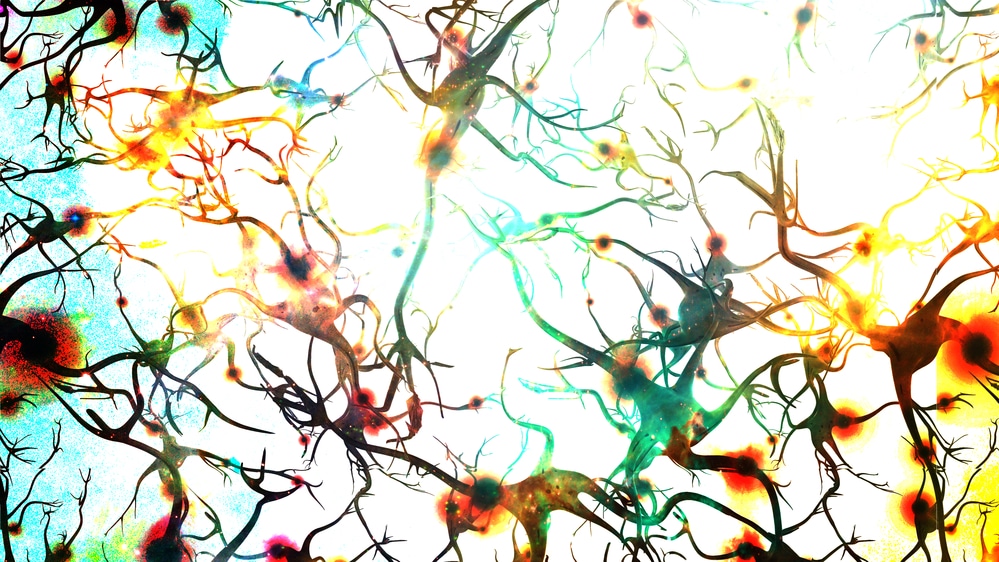
In a newly published study in the journal Scientific Reports, UIC researchers analyzed gene expression in fresh brain tissue. The study, entitled “Selective time-dependent changes in activity and cell-specific gene expression in human postmortem brain,” involved brain tissue that was first collected during routine brain surgery, then placed in an environment meant to simulate the “postmortem interval.” The findings were staggering. According to the study, researchers found that gene expression in some cells actually increased after death. This created a bit of a zombie-like effect. These so-called “zombie cells” were specific to one type of cell: inflammatory brain cells called glial cells. The researchers found that glial cells don’t just keep functioning after death; they actually grow and sprout long “arm-like appendages.”
Gene Stabilization After Death
In an attempt to understand the postmortem behavior of glial cells, researchers also studied a series of human genes. The researchers found that about 80 percent of the genes studied remained relatively stable for 24 hours after death; however, as expected, genes involved in human brain activity like memory and thinking rapidly degraded in the postmortem period. But even as the function of those genes degraded, the activity of another group increased. This third group of genes – again, cheekily called “zombie genes” – help support the glial cells. This helps explain the function of brain cells like glial cells after death.
Postmortem Gene Expression and Neurological Disorders
In a news release, researcher Dr. Jeffrey Loeb explained that the postmortem behavior of glial cells isn’t particularly surprising. “They are inflammatory and their job is to clean things up after brain injuries like oxygen deprivation or stroke,” Loeb said. However, the implications of the findings could be profoundly important to the study of neurological disorders. As Science Daily explains, most research that uses postmortem human brain tissues to study neurological disorders does not account for postmortem cell activity. “Most studies assume that everything in the brain stops when the heart stops beating, but this is not so,” Loeb said. This could revolutionize the study of neurological disorders ranging from autism spectrum disorders to Alzheimer’s disease. “Our findings don’t mean that we should throw away human tissue research programs, it just means that researchers need to take into account these genetic and cellular changes, and reduce the postmortem interval as much as possible to reduce the magnitude of these changes,” Loeb said.
_____
As expected, the behavior of brain cells like glial cells in the postmortem period has far-reaching consequences. This groundbreaking understanding of gene and cell behavior can inform the study of neurological conditions. From Alzheimer’s disease to autism spectrum disorders, the study could revolutionize future treatments.
Scantox is a part of Scantox, a GLP/GCP-compliant contract research organization (CRO) delivering the highest grade of Discovery, Regulatory Toxicology and CMC/Analytical services since 1977. Scantox focuses on preclinical studies related to central nervous system (CNS) diseases, rare diseases, and mental disorders. With highly predictive disease models available on site and unparalleled preclinical experience, Scantox can handle most CNS drug development needs for biopharmaceutical companies of all sizes. For more information about Scantox, visit www.scantox.com.


