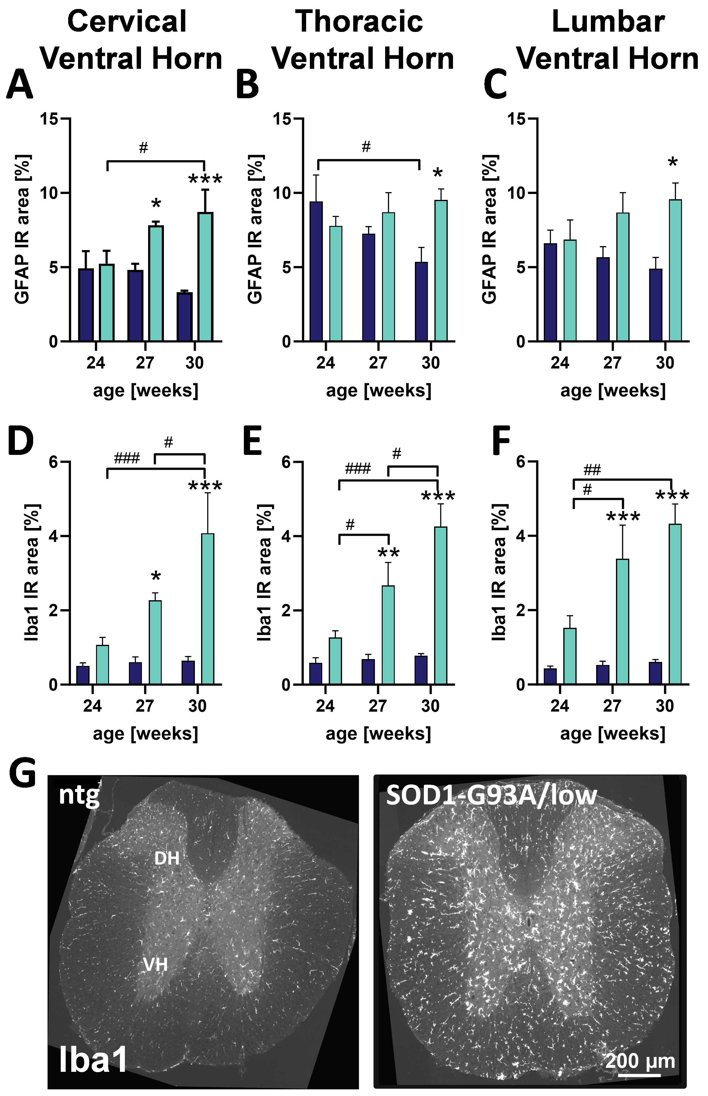
Amyotrophic Lateral Sclerosis (ALS) as progressive motor neuron disease is characterized by quickly progressing muscle atrophy and correlating motor symptoms. Degeneration of the upper and lower motor neurons of the spinal cord is the main cause for the observed motor decline and paralleled by strong neuroinflammation as observed by occurrence of astrocytosis and many activated microglia. The SOD1-G93A/low transgenic mouse as preclinical model of ALS expresses SOD1 with G93A mutation with a lower gene copy number compared to the commonly used SOD1-G93A model, causing a delayed symptom onset and extended survival and thus providing a longer window for drug treatment studies. Here we show, that male SOD1-G93A/low mice develop astrocytosis in the cervical, thoracic and lumbar ventral horn of the spinal cord as early as 27 weeks of age (GFAP; Fig.1A-C). Changes in microgliosis levels, as determined by an increased number of activated microglia, are even more pronounced than changes in astrogliosis in the same spinal cord regions. Additionally, activated microglia show a strong progression compared to non-transgenic (ntg) littermates (Iba1; Fig.1D-F).

Figure 1. Neuroinflammation in the spinal cord of male SOD1-G93A/low mice. Astrocytosis as analyzed by percent of cervical, thoracic and lumbar ventral horn immunoreactive (IR) area covered by GFAP (A-C) and activated microglia as analyzed by Iba1 IR area in the same spinal cord regions (D-F) in 24, 27 and 30 week old male SOD1-G93A/low mice. G: Representative images of Iba1 labeling in the cervical spinal cord of a 30 week old male SOD1-G93A/low mice compared to a ntg littermate. Two way ANOVA with Tukey’s and Sidak’s multiple comparison post hoc test. Mean + SEM. *p<0.05, **p<0.01, ***p<0.001. *differences between genotypes; # differences between age groups. DH: dorsal horn; VH: ventral horn.
Contact us today to get your study in SOD1-G93A/low mice started!


