Tau protein stabilizes microtubules and contributes to key structural and regulatory cellular functions as axonal transport and signaling. Neurofibrillary tangles, mainly composed of bundles of tau are implicated in the pathogenesis of neurodegenerative diseases such as tauopathies including Alzheimer‘s disease.
To identify inhibitors of tau aggregation Scantox provides an cell free appraoch as high trough screening tool.
This approach is a spectrofluorometric read out that can be performed as tau aggregation or dis-aggregation assay. The tau aggregation assay aims to identify compounds that are able to mitigate the aggregation of tau. The tau dis-aggregation assay investigates if developmental compounds are able to reverse tau aggregation.
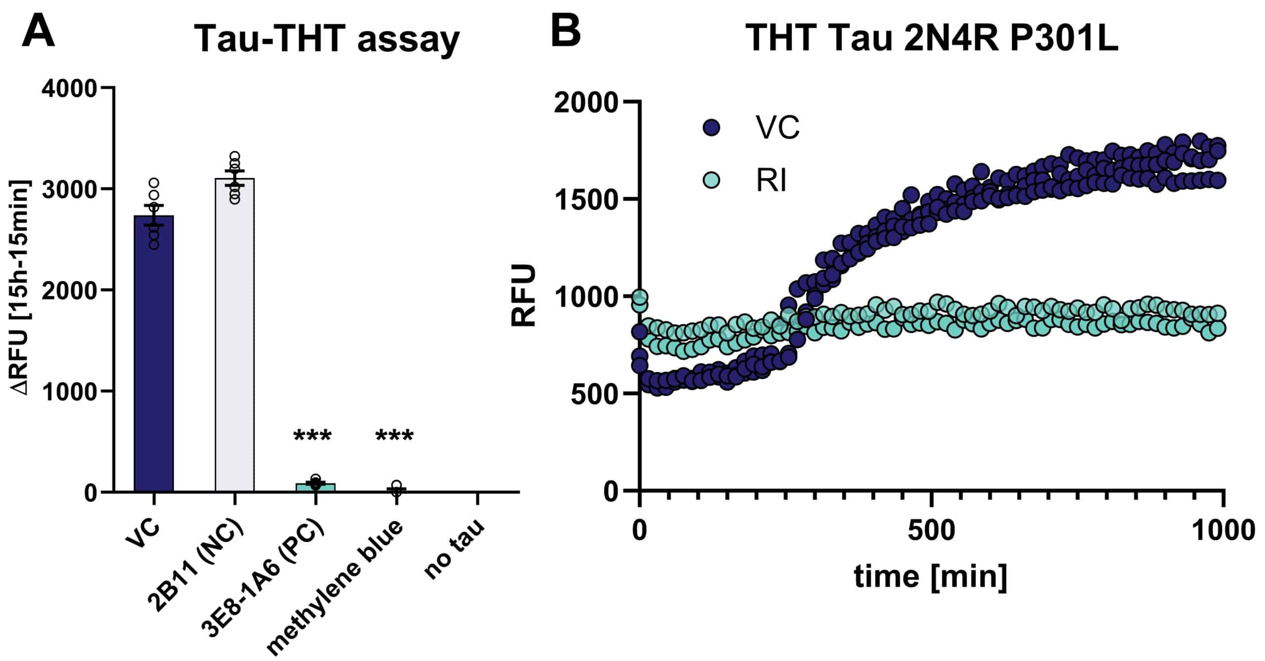
These assays are cell-free in vitro assays using recombinant Tau441 (2N4R) P301L that is incubated in an aggregation buffer including ThioS. Fluorescence intensity is detected at 465 nm excitation and 510 nm emission and can be monitored over time to additionally investigate kinetics of Tau aggregation (Quint et al., 2021).

Figure 1: Cell free THT-aggregation assay using recombinant 2N4R tau with P301L mutation. A: Results of aggregation assay shown as ΔRFU values. The antibodies 2B11 (negative control NC, specific for phoyphorylated tau), 3E8-1A6 (positive control PC, targeting microtubule binding domain) as well as methylene blue as inhibitor of aggregation were assessed. One-Way ANOVA with Dunnett’s multiple comparisons test vs. vehicle control (VC). Mean ± SEM; n = 6. ***p<0.001. B: monitoring tau aggregation over time using the THT assay; blue = vehicle control (VC), turquoise (RI) = antibody 3E8-1A6 as aggregation inhibitor.
