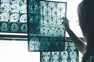
Three-dimensional (3D) cell culture systems are rapidly emerging as indispensable tools for modeling human disease and supporting drug discovery. Next to brain organoids, neural spheroids—spherical aggregates of progenitor or differentiated cells—offer reproducibility, scalability, and relatively short cultivation times. These features make them particularly well-suited for capturing cellular events and recapitulating pathological hallmarks of neurodegenerative disorders in vitro. In alignment with FDA guidance on New Approach Methodologies (NAMs), Scantox Neuro has invested substantial R&D efforts into establishing and characterizing human-relevant 3D models. One approach is based on iCell NeuroSpheres (FUJIFILM Cellular Dynamics), where we have validated fundamental characteristics such as neuronal activity and cell distribution, providing a robust foundation for disease modelling (Figure 1).

Figure 1: Baseline characterization of neurospheres. Cell distribution (A, B) of either PFC-like (A) or VTA-like (B) Neurospheres. Neurospheres were fixed on DIV21 and subject to whole-sphere immunocytochemical straining and confocal imaging (PFC: blue = Hoechst (Nuclei), turquoise = MAP2 (all neurons), yellow= GFAP (astrocytes) VTA blue = Hoechst (Nuclei), turquoise = MAP2 (all neurons), magenta = TH (dopaminergic neurons), yellow = GFAP). Neuronal activity (C, D) indirectly visualized with a genetically encoded calcium indicator (GECI) on vehicle treated PFC-like (A) or VTA-like (B) Neurospheres.
Alzheimer’s Disease (AD) Modelling
- Prefrontal cortex-like spheroids composed of glutamatergic and GABAergic neurons, along with astrocytes, were challenged with aggregated recombinant Aβ1-42
- Pathological markers, including Aβ deposition and astrocyte activation (GFAP), were visualized using advanced 3D confocal imaging (Figure 2)

Figure 2: Immunostaining of PFC-like iCell NeuroSpheres. iCell Neurospheres (FUJIFILM Cellular Dynamics) were cultured and treated with Aβ1-42 for 72 hours. Then, spheroids were fixed and stained with 6E10, MAP2 and GFAP markers. Z-stack images were obtained with ImageXpress HT.ai confocal microscope (Molecular devices).
Parkinson’s Disease (PD) Modelling
- Ventral tegmental area-like spheroids, incorporating dopaminergic neurons aside of glutamatergic and GABAergic neurons as well as astrocytes, were subjected to two lesion paradigms:
- Genetic insult via AAV-mediated expression of α-synuclein (A53T mutation)
- Chemical challenge with the neurotoxin MPP+
- Dopaminergic neuron integrity was assessed by immunocytochemistry (Figure 3), while mitochondrial function was evaluated using TMRM live-cell assays (Figure 4)

Figure 3: Dopaminergic neurons loss of VTA-like iCell NeuroSpheres after treatment. A: VTA-like iCell Neurospheres (FUJIFILM Cellular Dynamics) were treated with MPP+ for 48 hours. Dopaminergic loss was determined as (B) a reduction of TH intensity assessed with ImageXpress HT.ai confocal microscope (Molecular devices). Mean + SEM; n = 3-5, One-way ANOVA followed by Dunnet´s multiple comparisons test compared to vehicle control (VC). **p <0.01.

Figure 4: Mitochondrial activity in VTA-like iCell NeuroSpheres was evaluated by TMRM assay using Incucyte®. iCell NeuroSpheres were treated with MPP+, AAV-A53T, or both for 48 h. A: Total TMRM intensity over time. B: Total TMRM intensity at 48 h. Mean + SEM; n = 5. One-way ANOVA followed by Dunnet’s multiple comparison test compared to vehicle control (VC). ***p <0.001. OCU: Total orange object intensity.
Expanding the Platform
The integration of additional cell types (e.g., microglia, oligodendrocytes) and diverse lesion paradigms or genetic modifications further enhances the translational relevance of these models. Ongoing work at Scantox Neuro focuses on refining these systems to better capture disease complexity and accelerate therapeutic discovery.
Conclusion
Neural spheroid models represent a reproducible and scalable platform for investigating cellular alterations underlying human neurodegenerative pathophysiology. By bridging the gap between conventional preclinical models and patient-relevant biology, these systems provide a powerful foundation for drug discovery and translational research.
Our mission: to provide scalable, reproducible platforms that bridge the gap between preclinical research and patient-relevant therapies. We welcome collaborations to explore new applications together.